
衡道病理特邀撰稿作者翻译了WHO部分图⽚的图注,并用手帐的⽅式进行中英⽂对照,且通过不同颜色的划线将晦涩难懂的英文单词与中⽂翻译同时标注,希望对专业英文的学习有所帮助。
由于本篇目的以英文学习为主,篇幅有限,故未对各个疾病进行详细阐述。全部图片均来自WHO,若有不恰当之处,还请评论区指正。
Serous cystadenoma, adenofibrom a , and surface papilloma of the ovary 卵巢浆液性囊腺瘤 , 腺纤维瘤和表面乳头状瘤
Definition
Serous cystadenoma, adenofibroma, and surface papilloma are benign serous tumours composed of cells resembling fallopian tube epithelium.
定义
浆液性囊腺瘤、腺纤维瘤和表⾯乳头状瘤是由类似输卵管上⽪细胞组成的良性浆液性肿瘤
Essential and desirable diagnostic criteria
Essential: tumour with benign serous epithelium, with or without fibromatous stroma; for cystadenoma, the cysts should be >1 cm
基本的诊断标准
基本特征:良性浆液上皮肿瘤,伴或不伴纤维瘤间质;囊腺瘤,囊肿大小应为>1cm。
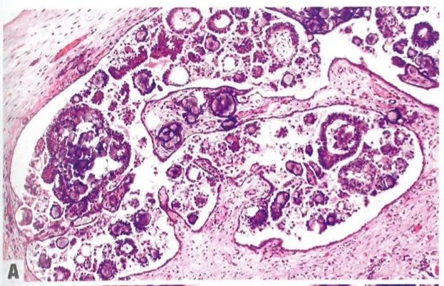
Fig. 1.04 A Ovarian serous cystadenofibroma.
The tumour is predominantly cystic and the cyst lining is mostly smooth, with some nodular areas.
B Serous cystadenofibroma.
Glands lined by bland tubal-type epithelium, with occasional ciliated cells, are present in a fibromatous stroma.
图1.04 A卵巢浆液性囊腺纤维瘤。肿瘤主要是囊性的,囊壁⼤部分平滑,有⼀些结节状区域。
B浆液性囊腺纤维瘤。在纤维间质中,有内衬温和的输卵管型上⽪的腺体,偶有纤⽑细胞。
Fig. 1.05 Serous surface papilloma. Stromal papillae covered by bland tubal-type epithelium project from the ovarian surface.
图1.05浆液性表⾯乳头状瘤。被覆输卵管型上皮的乳头,从卵巢表面突出。
Serousborderline tumour of the ovary
卵巢浆液性交界性肿瘤
Definition
Serous borderline tumour (SBT) is a non-invasive, low-grade, proliferative serous epithelial neoplasm.
定义
浆液性交界性肿瘤(SBT)是⼀种非浸润性、低级别、增生性浆液性上皮肿瘤。
Essential and desirable diagnostic criteria SBT
Essential: epithelial proliferation (with papillary hierarchical branching or micropapillary/cribriform pattern, low-grade cytology) with no stromal invasion. SBT with microinvasion Essential: stromal microinvasion < 5 mm in greatest dimension in any single focus with small cell clusters / individual cells with abundant dense eosinophilic cytoplasm or small papillae within clear lacunar spaces, cytologically similar to the noninvasive component of SBT.
基本的诊断标准
SBT
基本特征:上皮细胞增生(伴有多级乳头状分或微乳头状/筛状模式,低级 别细胞学特征),无间质浸润。SBT伴微浸润 基本特征:在任何单个病灶中,间质微浸润最⼤径< 5 mm,微浸润灶为间 质内强嗜酸性细胞质的⼩细胞簇/单个细胞,或收缩间隙内小乳头。细胞学上类似于SBT的⾮浸润性成分。
Fig. 1.06 Serous borderline tumour.
A Papillae are architecturally complex, with hierarchical branching.
B Papillae are lined by stratifified epithelium with detached small clusters of tumour cells.
图1.06交界性浆液性肿瘤。
A 乳头在结构上是复杂的,具有多级分支。
B 乳头由复层上⽪和脱落的小簇肿瘤细胞构成。
Fig. 1.07 Micropapillary serous borderline tumouir.
A Micropapillary architecture is prominent.
B Elongated micropapillae directly emanate from a large papilla, producing the so-called Medusa head appearance.
图1.07微乳头状浆液性交界性肿瘤。
A 明显的微乳头状结构。
B 细⻓的微乳头直接从⼀个⼤乳头发出,产⽣所谓的美杜莎头外观。
Fig. 1.08 Epithelial-type non-invasive implants associated with ovarian serous borderline tumour.
A Complex papillary architecture is present within an epithelium-lined space.
B Papillae contain cells with low-grade nuclear features. Psammoma bodies are present.
图1.08与卵巢浆液性交界性肿瘤相关的上皮性⾮浸润性种植。
A 在上⽪内衬的间隙内有复杂的乳头状结构。
B 乳头含有低级别核特征的细胞。有砂砾体。
Fig. 1.09 Desmoplastic non-invasive implants associated with ovarian serous borderline tumour.
A .Low-grade simple glands with slight epithelial stratifification are present within abundant desmoplastic stroma.
B .Individual epithelial cells (arrows) are present within abundant desmoplastic stroma. This fifinding does not fulfifil the criteria for invasive implants.
图1.09 与卵巢浆液性交界性肿瘤相关的促结缔组织增⽣性⾮浸润性种植。
A .在丰富的结缔组织间质中存在低 级别简单腺体伴轻度复层上⽪。
B .单个上⽪细胞(箭头)位于丰富的 促结缔组织间质中。这⼀发现不符合浸润性种植的标准。
Fig.1.10 Invasive implants associated with ovarian serous borderline tumour.
AThere is destructive infifiltration of underlying tissue. Small papillae and micropapillae are markedly crowded and haphazardly arranged. The histological appearance is akin to that of invasive low-grade serous carcinoma.
BSmall papillae and micropapillae are present within clear lacunar spaces and demonstrate low-grade atypia. Psammoma bodies are also seen.
图1.10卵巢浆液性交界性肿瘤相关的浸润性种植。
A组织有破坏性浸润。⼩乳头和微乳头明显拥挤和无序排列。组织学外观类似于浸润性低级别浆液性癌。
B收缩的裂隙内可见小乳头和微乳头,表现为轻度不典型性。也可以看到砂砾体。
Fig. 1.11 Microinvasion in a serous borderline tumour.
Small nests and individual tumour cells with abundant eosinophilic cytoplasm (arrows) are present within spaces devoid of epithelial lining in the stroma. This is the most common pattern of microinvasion.
图1.11浆液性交界性肿瘤伴微浸润。
可见小巢状肿瘤细胞和富含嗜酸性细胞质的单个肿瘤细胞(箭头),周围存 在收缩间隙。这是最常见的微浸润模式。
Fig. 1.12 Microinvasive low-grade serous carcinoma.
Crowded small and medium-sized papillae in clear lacunar spaces (< 5 mm in greatest extent) are similar to the morphology of an invasive low-grade serous carcinoma.
图1.12微浸润性低级别浆液性癌。收缩腔隙内密集的中小型乳头突起(最⼤直径< 5mm),与浸润性低级别浆液性癌的形态相似。
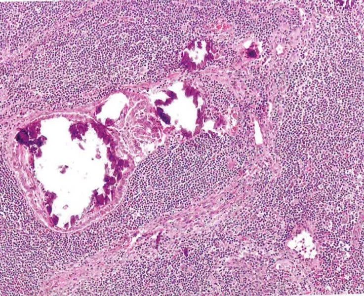
Fig. 1.13 Serous borderline tumour in a pelvic lymph node. Papillary proliferation of bland serous epithelium and psammoma bodies, at the left. There is also endosalpingiosis involving this node, at the right.
图1.13盆腔淋巴结中的浆液性交界性肿瘤。左侧为淡染的浆液性上皮呈乳头状增生和沙砾体。右侧也可⻅累及此淋巴结的输卵管上⽪异位。
Fig. 1.14 Endosalpingiosis. Epithelial stratifification, cell tufting/detachment, and papillary architecture of epithelial-type non-invasive implants are absent. Endosalpingiosis does not qualify as advanced-stage in the setting of an ovarian serous borderline tumour.
图1.14 输卵管内膜异位症。缺乏上⽪细胞复层、成簇/脱落和乳头状结构的非浸润性种植。在卵巢浆液性交界性肿瘤的背景下,输卵管内膜异位症不符合晚期标准。
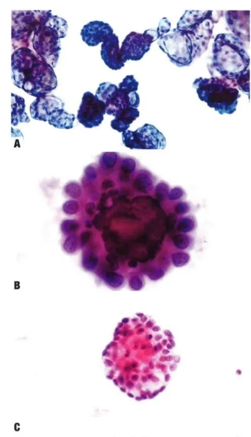
Fig. 1.15 A Low-grade serous neoplasm.
A Papillary structures with relatively uniform small cells and low-grade nuclear atypia.
B Serous borderline tumour. Peritoneal flfluid cytology. Note papillae with mild epithelial atypia encompassing a psammoma body.
C Serous borderline tumour. Peritoneal flfluid cytology. Note the 3D epithelial clusters with mild epithelial atypia and discernible nucleoli.
图1.15低级别浆液性肿瘤。
A乳头状结构,相对均匀的小细胞和低 级别核异型性。
B浆液性交界性肿瘤。腹水细胞学。可见乳头状结构,衬覆轻度异型上皮,包围砂砾体。C浆液性交界性肿瘤。腹水细胞学。可见3D上⽪簇,上⽪呈轻度异型,可见核仁。
Low-grade serous carcinoma of the ovary
卵巢低级别浆液性癌
Definition
Low-grade serous carcinoma (LGSC) is an invasive serous neoplasm with low-grade malignant features.
定义
低级别浆液性癌是⼀种具有低级别恶性特征的浸润性浆液性肿瘤。
Essential and desirable diagnostic criteria
Essential: invasive serous tumour with small nests, glands, papillae or micropapillae, and inverted macropapillae, frequently free-floating within unlined clear spaces; low-grade cytological atypia (< 3-fold variation in nuclear size); low mitotic activity.
基本的诊断标准
基本特征:浸润性浆液性肿瘤,有小巢、腺样、乳头或微乳头和倒置大乳头,常在无衬覆上皮的收缩间隙内自由漂浮;低级别细胞异型性(核大小< 3倍);低核分裂计数。
Fig. 1.16 Low-grade serous carcinoma. A large multicystic mass with nodular areas and excrescences.
图1.16低级别浆液性癌。大的多囊肿块,有结节状区域和赘生物。
Fig.1.17 Low-grade serous carcinoma. Small papillae containing cells with uniform nuclei and inconspicuous mitotic activity.
图1.17低级别浆液性癌。小乳头含有均匀⼀致的细胞核和不明显的核分裂象。
Fig. 1.18 Low-grade serous carcinoma. This example exhibits an inverted macropapillary pattern of invasion. The macropapillae are surrounded by unlined clear spaces.
Scattered small papillae, clusters, and individual cells are also present.
图1.18低级别浆液性癌。图示倒置大乳头状浸润模式。大乳头被无上皮衬覆的收缩间隙包围。也可见散在的小乳头、簇状和单个细胞。
Fig.1.19 Low-grade serous carcinoma.
This peritoneal washing specimen shows numerous papillary clusters of uniform cells, as well as psammoma bodies. Note that the fifindings in this specimen are not diagnostic of low-grade serous carcinoma and could also be seen in association with a serous borderline tumour .
图1.19低级别浆液性癌。腹膜冲洗标本可见大量均匀的乳头状细胞簇,以及砂砾体。需要注意的是,本标本中的发现不能诊断低级别浆液性癌,也可见于浆液性交界性肿瘤。
High-grade serous carcinoma of the ovary
卵巢⾼级别浆液性癌
Definition
High-grade serous carcinoma (HGSC) is a high-grade epithelial neoplasm demonstrating serous differentiation.
定义
高级别浆液性癌(HGSC)是⼀种显示浆液性分化的高级别上皮肿瘤。
Essential and desirable diagnostic criteria
Essential: serous tumour with solid (with slit-likespaces), papillary, glandular, or cribriform ar chitecture; large, markedly atypical nuclei (nuclear size variability of > 3-fold); high mitotic activity. Desirable (in selected cases): WT1 immunoreactivity; mutationtype p53 expression.
基本的诊断标准
基本特征:浆液性肿瘤,呈实性(伴裂隙样间隙)、乳头状、腺状或筛状结 构;⼤⽽明显的异型性核(核⼤⼩ >正常核3倍);高核分裂计数。辅助诊断(免疫组化):WT1阳性;p53呈突变型表达。
Fig. 1.20 High-grade serous carcinoma. Papillary structures containing cells with markedly pleomorphic nuclei and conspicuous mitotic activity.
图1.20⾼级别浆液性癌。具有 明显多形性核和较多核分裂的细胞构成乳头状结构。
Fig.1.21 High-grade serous carcinoma.
A Papillary architecture.
B Solid architecture with tumour-infifiltrating lymphocytes.
C Prominent clear cell change, mimicking clear cell carcinoma. This fifinding is often focal, and the clear cell areas show the same immunophenotype as conventional high-grade serous carcinoma.
D Intracytoplasmic mucin in some tumour cells .
图1.21⾼级别浆液性癌。
A乳头状结构。
B肿瘤浸润淋巴细胞的实性结构。
C透明细胞改变明显,类似透明细胞癌。这⼀发现通常是局灶的,透明细胞区域显示与经典型高级别浆液性癌相同的免疫表型。
D部分肿瘤细胞胞浆内黏蛋白(黏蛋白染⾊阳性)。
Fig. 1.22 High-grade serous carcinoma (HGSC).
A SET (solid, endometrial-like, transitional) pattern of HGSC with glandular (pseudoendometrioid) architecture.
B SET pattern of HGSC with transitional cell-like architecture.
C Diffffuse WT1 expression in an HGSC with solid and pseudoendometrioid (cribriform) architecture.
图1.22⾼级别浆液癌(HGSC)。
A SET(实性,子宫内膜样,移行细胞样)型HGSC伴腺样(假子宫内膜样)结 构。
B 具有移⾏细胞样结构的HGSC SET模式。
C WT1在实性和假⼦宫内膜样(筛状)结构的HGSC中弥漫表达。
Fig. 1.23 High-grade serous carcinoma.
A Mutant-pattern p53 immunostaining with strong nuclear expression in almost all tumour cell nuclei (> 80%).
B Mutant-pattern p53 immunostaining with complete loss of expression in tumour cells. Note that the retained staining in benign cells serves as an internal control.
C Mutant-pattern p53 immunostaining with variable cytoplasmic staining. This is the least common staining pattern seen in highgrade serous carcinoma.
图1.23⾼级别浆液性癌。
A突变型p53免疫染色,几乎所有肿瘤细胞核弥漫强阳性(> 80%)。
B突变型p53免疫染色,在肿瘤细胞中完全失表达。注意,良性细胞中保留的染色作为内对照。
C突变型p53免疫染色,不同程度的胞浆染色。这是高级别浆液性癌中最不常见的染色模式。
Fig.1.24 High-grade serous carcinoma. Pleomorphic tumour cells with large plasmic vacuoles and marked nuclear atypia .
图1.24高级别浆液性癌。多形性肿瘤细胞具有⼤的胞浆内空泡和明显的核异型性。
整理:南瓜子儿
审核:樊月
设计:鹏飞
编辑:V
本公众号发布所有原创内容,版权均属衡道医学病理诊断中心及相关版权方所有,内容仅供学术交流,如有侵权请联系删除。未经授权的转载是侵权行为,版权方保留追究法律责任的权利。投稿、转载或版权疑议,请联系:tougao@histo.cn;
不感兴趣
看过了
取消

人点赞

人收藏

打赏
不感兴趣
看过了
取消
©2012-2023 北京华媒康讯信息技术股份有限公司 All Rights Reserved. 注册地址:北京 联系电话:010-82736610
广播电视节目制作经营许可证 —(京)字第 17437号 京海食药监械经营备20200522号
京ICP备12011723号 京ICP证150092号
 京公网安备 11010802020745号
工商备案公示信息
互联网药品信息服务资格证书((京)-非经营性-2020-0015)
京公网安备 11010802020745号
工商备案公示信息
互联网药品信息服务资格证书((京)-非经营性-2020-0015)


您已认证成功,可享专属会员优惠,买1年送3个月!
开通会员,资料、课程、直播、报告等海量内容免费看!

打赏金额
认可我就打赏我~
1元 5元 10元 20元 50元 其它
打赏作者
认可我就打赏我~
扫描二维码
立即打赏给Ta吧!
温馨提示:仅支持微信支付!
已收到您的咨询诉求 我们会尽快联系您


 010-82736610
 股票代码: 872612


